Computed tomograph and digital angiograph in the evaluation of coarctation of the aorta: Experience from a pediatric hospital in Peru, 2013-2023
DOI:
https://doi.org/10.58597/rpe.v4i3.119Keywords:
Coarctation of the aorta, Helical Computed Tomography, Digital Subtraction Angiography, Congenital heart diseaseAbstract
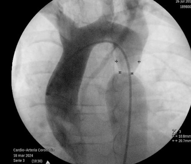
Objective: To compare aortic vessel measurements obtained by CT and DA in patients diagnosed with aortic coarctation treated at a pediatric hospital in Peru. Materials and methods: An observational, descriptive, and cross-sectional study was conducted through a retrospective review of medical records of patients under 18 years of age diagnosed with CoA and treated between 2013 and 2023. Epidemiological, clinical, and aortic vessel measurement variables were collected, and diameters obtained by both techniques were compared. Results: Of the 42 patients included in the study, 64.3% were male, with a median age of 8.5 years (IQR: 3–13.25). The postductal form was the most frequent (52,3%). Therapeutic catheterization was the predominant treatment (64%). In the comparison of aortic measurements, DA showed slightly higher values in the aortic arch, isthmus, and coarctation site, although no statistically significant differences were found compared to CT (p>0.05). Conclusion: CT and DA showed similar measurements in the evaluation of aortic anatomy in pediatric patients with CoAo. In Peru, where access to invasive procedures is centralized, CT is a highly valuable noninvasive diagnostic tool for anatomical characterization and follow-up, particularly when immediate therapeutic intervention is not required.
Downloads
References
Raza S, Aggarwal S, Jenkins P, Kharabish A, Anwer S, Cullington D, Jones J, Dua J, Papaioannou V, Ashrafi R, Moharem-Elgamal S. Coarctation of the Aorta: Diagnosis and Management. Diagnostics (Basel). 2023;13(13):2189. doi: 10.3390/diagnostics13132189.
Turaev BB, Abralov KK, Kobiljonov BK, et al. Assessment of anatomy of the aorta in patients with a coarctation of aorta. Cardiothorac Surg. 2023;31:24. doi:10.1186/s43057-023- 00114-w
Grattan M., Prince A., Rumman R.K., Morgan C., Petrovic M., Hauck A., Young L., Franco-Cereceda A., Loeys B., Mohamed S.A., et al. Predictors of Bicuspid Aortic Valve–Associated Aortopathy in Childhood. Circ. Cardiovasc. Imaging. 2020;13:e009717. doi: 10.1161/CIRCIMAGING.119.009717
Browne LP, Barker AJ, Vargas D. Imaging Follow-up of Repaired Aortic Coarctation. Semin Roentgenol. 2020;55(3):301-311. doi: 10.1053/j.ro.2020.06.011.
Bolaños I, Mora K, Bolaños S, Bujan S. Coartación de aorta. Med. leg. Costa Rica. 2020; 37(1): 87-92. http:// www.scielo.sa.cr/scielo.php?script=sci_arttext&pid=S1409- 00152020000100087&lng=en.
Lucheniuc C, Layerle B, Pujadas M, Chiesa P, Pírez M, Alegretti
M. Coartación de aorta en pediatría: características clínicas de niños y adolescentes asistidos en el Centro Hospitalario Pereira Rossell. Rev.Urug.Cardiol. 2023; 38(1): e203. https://doi. org/10.29277/cardio.38.1.12.
Salciccioli KB, Zachariah JP. Coarctation of the Aorta: Modern ParadigmsAcrossthe Lifespan. Hypertension. 2023; 80(10):1970- 1979. doi:10.1161/HYPERTENSIONAHA.123.19454
Pinto Júnior VC, Branco KM, Cavalcante RC, Carvalho Junior W, Lima JR, Freitas SM, Fraga MN, Souza NM. Epidemiology of congenital heart disease in Brazil. Rev Bras Cir Cardiovasc. 2015;30(2):219-24. doi: 10.5935/1678-9741.20150018.
Ardila DF, Rincón-Montana AG, García-Pérez LD, Gracia G, Zarante I. Prevalencia de coartación aórtica en Bogotá- Colombia de 2001 a 2018. El diagnóstico debe mejorar [Prevalence of aortic coarctation in Bogotá-Colombia from 2001 to 2018. The diagnostic needs to improve]. Arch Cardiol Mex. 2022;92(2):196-202. doi: 10.24875/ACM.20000515.
Torres C, Uriondo V, Ramirez A, Arroyo H, Loo M, Protzel A, et al. Factores asociados a la supervivencia al año de vida en neonatos con cardiopatía congénita severa en un Hospital Nacional del Perú. Rev Peru Med Exp Salud Publica. 2019; 36( 3 ): 433-441. doi: 10.17843/rpmesp.2019.363.4166.
Yokoyama U, Ichikawa Y, Minamisawa S, Ishikawa Y. Pathology and molecular mechanisms of coarctation of the aorta and its association with the ductus arteriosus. J Physiol Sci. 2017;67(2):259-270. doi: 10.1007/s12576-016-0512-x.
Dijkema EJ, Leiner T, Grotenhuis HB. Diagnosis, imaging and clinical management of aortic coarctation. Heart. 2017 Aug;103(15):1148-1155. doi: 10.1136/heartjnl-2017-311173.
Epub 2017 Apr 4. Erratum in: Heart. 2019;105(14):e6. doi: 10.1136/heartjnl-2017-311173corr1.
Gong T, Zhang F, Feng L, Zhu X, Deng D, Ran T, Li L, Kong L, Sun L, Ji X. Diagnosis and surgical outcomes of coarctation of the aorta in pediatric patients: a retrospective study. Front Cardiovasc Med. 2023;10:1078038. doi: 10.3389/ fcvm.2023.1078038.
Huang F, Chen Q, Huang WH, Wu H, Li WC, Lai QQ. Diagnosis of Congenital Coarctation of the Aorta and Accompany Malformations in Infants by Multi-Detector Computed Tomography Angiography and Transthoracic Echocardiography: A Chinese Clinical Study. Med Sci Monit. 2017;23:2308-2314. doi: 10.12659/msm.901928.
Familiari A, Morlando M, Khalil A, Sonesson SE, Scala C, Rizzo G, Del Sordo G, Vassallo C, Elena Flacco M, Manzoli L, Lanzone A, Scambia G, Acharya G, D’Antonio F. Risk Factors for Coarctation of the Aorta on Prenatal Ultrasound: A Systematic Review and Meta-Analysis. Circulation. 2017;135(8):772-785. doi: 10.1161/CIRCULATIONAHA.116.024068.
Goudar SP, Shah SS, Shirali GS. Echocardiography of coarctation of the aorta, aortic arch hypoplasia, and arch interruption: strategies for evaluation of the aortic arch. Cardiol Young. 2016;26(8):1553-1562. doi: 10.1017/S1047951116001670.
Miabi Z, Pourfathi H, Midia M, Midia R, Parvizi R. Comparison of CT angiography and digital subtraction angiography in the diagnosis of aortic coarctation. Pak J Biol Sci. 2011;14(1):74-7. doi: 10.3923/pjbs.2011.74.77.
Leo I, Sabatino J, Avesani M, Moscatelli S, Bianco F, Borrelli N, De Sarro R, Leonardi B, Calcaterra G, Surkova E, Di Salvo G, On Behalf Of The Working Group On Congenital Heart Disease Cardiovascular Prevention In Paediatric Age Of The Italian Society Of Cardiology Sic. Non-Invasive Imaging Assessment in Patients with Aortic Coarctation: A Contemporary Review. J Clin Med. 2023;13(1):28. doi: 10.3390/jcm13010028.
Krylova A, Svobodov A, Tumanyan M, Levchenko E, Kotov S, Butrim Y, Shvartz V. Results of Aortic Coarctation Repair in Low- and Normal Birth-Weight Neonates: A Propensity Score-Matched Analysis. Life (Basel). 2023;13(12):2282. doi: 10.3390/life13122282.
Sezer SS, Narin N, Ozyurt A, Onan SH, Pamukcu O, Argun M, Baykan A, Uzum K. Cardiovascular changes in children with coarctation of the aorta treated by endovascular stenting. J Hum Hypertens. 2014;28(6):372-7. doi: 10.1038/jhh.2013.119.
Doroshenko OV, Kuchumov AG, Golub MV, Rakisheva IO, Skripka NA, Pavlov SP, Strazhec YA, Lazarkov PV, Saychenko ND, Shekhmametyev RM. Investigation of Relationship between Hemodynamic and Morphometric Characteristics of Aortas in Pediatric Patients. J Clin Med. 2024;13(17):5141. doi: 10.3390/jcm13175141.
Xiao HJ, Zhan AL, Huang QW, Huang RG, Lin WH. Computed tomography angiography assessment of the degree of simple coarctation of the aorta and its relationship with surgical outcome: A retrospective analysis. Front Pediatr. 2022;10:1017455. doi: 10.3389/fped.2022.1017455.
Lucheniuc C, Layerle B, Pujadas M, Chiesa P, Pírez MC, Alegretti M. Coartación de aorta en pediatría: características clínicas de niños y adolescentes asistidos en el Centro Hospitalario Pereira Rossell. Rev Urug Cardiol. 2023;38(1):e12. doi:10.29277/ cardio.38.1.12
Gach P, Dabadie A, Sorensen C, Quarello E, Bonello B, Pico H, Hugues N, Petit P, Gorincour G. Multimodality imaging of aortic coarctation: From the fetus to the adolescent. Diagn Interv Imaging. 2016;97(5):581-90. doi: 10.1016/j.diii.2016.03.006.
Ganigara M, Doshi A, Naimi I, Mahadevaiah GP, Buddhe S, Chikkabyrappa SM. Preoperative Physiology, Imaging, and Management of Coarctation of Aorta in Children. Semin Cardiothorac Vasc Anesth. 2019;23(4):379-386. doi: 10.1177/1089253219873004. Epub 2019 Sep 19. Erratum in:
Semin Cardiothorac Vasc Anesth. 2021 Mar;25(1):77. doi: 10.1177/1089253220958571.
Bravo-Jaimes K, Lozano D, Orozco J, Rosales W, Macedo N, Medina M, et al. Tamizaje neonatal de cardiopatías congénitas críticas en el Perú: un llamado de urgencia. Arch Peru Cardiol Cir Cardiovasc. 2024;5(3):157-166. doi: 10.47487/apcyccv. v5i3.366
Tauma-Arrué A, Chávez-Saldivar S, Mego JC, Luis-Ybáñez O, Coronado-Quispe J, Lucena S, Alvarez C, Melgar E, Morales A, Marquez R, Wilhalme H, Bravo-Jaimes K. Trends in outpatient visits and deaths due to congenital heart defects in Peru. Int J Cardiol Congenit Heart Dis. 2022;7:100334. doi: 10.1016/j. ijcchd.2022.100334.
Cueva-Ortega, L, Montenegro-Castro DC. Características de las cardiopatías congénitas en infantes con síndrome de Down en un hospital de Chiclayo, Perú: Characteristics of congenital heart disease in infants with Down syndrome in a hospital in Chiclayo, Peru. Revista Experiencia En Medicina Del Hospital Regional Lambayeque. 2025; 10(4): 37–44. doi:10.37065/rem. v10i4.796
Zaki M, Hassan M. Aortic coarctation: evaluation with computed tomography angiography in pediatric patients. Med J Cairo Univ. 2015;83(2):63-70.
Malone LJ, Morin CE, Browne LP. Coronary computed tomography angiography in children. Pediatr Radiol. 2022 Dec;52(13):2498-2509. doi: 10.1007/s00247-021-05209-2.
Vaz A, Young LM, Couto RM, de Paula KRM, Fonseca EKUN. CT angiography versus clinical, echocardiographic, and invasive gradients in coarctation and recoarctation of the aorta. Ann Pediatr Cardiol. 2025;18(1):19-25. doi: 10.4103/apc. apc_221_24.
Starmans NL, Krings GJ, Molenschot MM, van der Stelt F, Breur JM. Three-dimensional rotational angiography in children with an aortic coarctation. Neth Heart J. 2016;24(11):666-74. doi: 10.1007/s12471-016-0899-2.
Merchak AA. Angiotomografía computada en pediatría: experiencia en un hospital pediátrico. Rev Chil Radiol. 2008;14(2):73-9
van der Stelt F, Siegerink SN, Krings GJ, Molenschot MMC, Breur JMPJ. Three-Dimensional Rotational Angiography in Pediatric Patients with Congenital Heart Disease: A Literature Review. Pediatr Cardiol. 2019 Feb;40(2):257-264. doi: 10.1007/ s00246-019-02052-z.
Downloads
Published
Issue
Section
License
Copyright (c) 2025 Diana C. Mucha-Lopez, Mimia Macuri-Valle, Carlos Mariño-Vigo

This work is licensed under a Creative Commons Attribution 4.0 International License.