Sociodemographic, clinical, and surgical characteristics of patients undergoing myelomeningocele repair at a public pediatric institute in Peru, 2017–2022
DOI:
https://doi.org/10.58597/rpe.v5i1.140Keywords:
Myelomeningocele, Infant, Newborn, Surgical Procedures, Operative, Postoperative ComplicationsAbstract
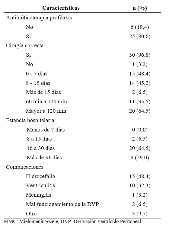
Objective: To describe the sociodemographic, clinical, and surgical characteristics of patients who underwent myelomeningocele repair at a public pediatric institute in Peru between 2017 and 2022. Materials and Methods: An observational, descriptive, cross-sectional, and retrospective study was conducted. Neonates diagnosed with myelomeningocele who underwent surgical repair at INSN Breña between January 2017 and December 2022 were included. Stillbirths, fetal deaths, and incomplete medical records were excluded. Maternal sociodemographic and obstetric variables, neonatal characteristics, and surgical treatment variables were collected. Descriptive analysis was performed using absolute and relative frequencies with Stata v.16 software. Results: Out of the 31 medical records, most patients were born at term (54.8%), with a birth weight between 2500 and 3500 g (71.0%), and via eutocic delivery (64.5%). The most frequent defect location was lumbosacral (74.2%). Hydrocephalus was present in 96.8% of cases. Prophylactic antibiotic therapy was administered in 80.6%, and 96.8% of surgeries were considered technically adequate. Surgical repair was performed within the first 15 days of life in 93.6% of patients. The most frequent complications were hydrocephalus, ventriculitis, and ventriculoperitoneal shunt dysfunction. Hospital stay exceeded 16 days in 93.5% of cases. Conclusion: Early myelomeningocele repair showed adequate technical outcomes; however, the high frequency of complications and prolonged hospital stay highlight the need for timely, standardized, and multidisciplinary clinical–surgical management to improve neonatal outcomes.
Downloads
References
Karsonovich T, Nethi S, Arya K. Meningocele. [Updated 2024 Oct 29]. In: StatPearls [Internet]. Treasure Island (FL): StatPearls Publishing; 2025 Jan-. Available from: https://www.ncbi.nlm.nih.gov/books/NBK562174/
Copp AJ, Adzick NS, Chitty LS, Fletcher JM, Holmbeck GN, Shaw GM. Spina bifida. Nat Rev Dis Primers. 2015;1:15007. doi: 10.1038/nrdp.2015.7.
Copp AJ, Greene ND. Neural tube defects--disorders of neurulation and related embryonic processes. Wiley Interdiscip Rev Dev Biol. 2013;2(2):213-27. doi: 10.1002/wdev.71.
Pattisapu JV, Veerappan VR, White C, Vijayasekhar MV, Tesfaye N, Rao BH, Park KB. Spina bifida management in low- and middle-income countries - a comprehensive policy approach. Childs Nerv Syst. 2023;39(7):1821-1829. doi: 10.1007/s00381-023-05988-z.
Alphonse B, Darko K, Michelande E, Limann B, Ovil R, Ulysse J, Barrie U, Detchou D, Lafortune Y, Valsaint JP. Myelomeningocele in the Pediatric Neurosurgery Department at Bernard Mevs Hospital in Haiti: A Retrospective Analysis. Neurosurg Pract. 2025;6(2):e00130. doi: 10.1227/neuprac.0000000000000130.
Montenegro J, Rodriguez S, Gonzalez I, Cortes E. Manejo Inicial de Mielomeningocele: Revisión de la Literatura: Manejo inicial de mielomeningocele. Neurocienc J. 2022;29(2):46-57. Disponible en: https://medcytjournals.com/index.php/neurocienciasjournal/article/view/391
Martinez H, Benavides-Lara A, Arynchyna-Smith A, Ghotme KA, Arabi M, Arynchyn A. Global strategies for the prevention of neural tube defects through the improvement of folate status in women of reproductive age. Childs Nerv Syst. 2023;39(7):1719-1736. doi: 10.1007/s00381-023-05913-4.
Ghotme KA, Arynchyna-Smith A, Maleknia P, Kancherla V, Pachon H, J Van der Wees P, Bocchino JM, Rosseau GL. Barriers and facilitators to the implementation of mandatory folate fortification as an evidence-based policy to prevent neural tube defects. Childs Nerv Syst. 2023;39(7):1805-1812. doi: 10.1007/s00381-023-05944-x.
Cavalheiro S, da Costa MDS, Moron AF, Leonard J. Comparison of Prenatal and Postnatal Management of Patients with Myelomeningocele. Neurosurg Clin N Am. 2017;28(3):439-448. doi: 10.1016/j.nec.2017.02.005.
Zoghi S, Feili M, Mosayebi MA, Ansari A, Feili A, Masoudi MS, Taheri R. Surgical outcomes of myelomeningocele repair: A 20-year experience from a single center in a middle-income country. Clin Neurol Neurosurg. 2024;239:108214. doi: 10.1016/j.clineuro.2024.108214.
Cehan AR, Dorobanțu DC, Tamas CI, Cehan VD, Tamas F, Balasa A. A Single-Centre Analysis of Surgical Techniques for Myelomeningocele Closure: Methods, Outcomes, and Complications. Clin Pract. 2024;14(5):2056-2070. doi: 10.3390/clinpract14050162.
Kesan K, Kothari P, Gupta R, Gupta A, Karkera P, Ranjan R, Mutkhedkar K, Sandlas G. Closure of large meningomyelocele wound defects with subcutaneous based pedicle flap with bilateral V-Y advancement: our experience and review of literature. Eur J Pediatr Surg. 2015;25(2):189-94. doi: 10.1055/s-0034-1368796.
Paslaru FG, Panaitescu AM, Iancu G, Veduta A, Gica N, Paslaru AC, Gheorghiu A, Peltecu G, Gorgan RM. Myelomeningocele Surgery over the 10 Years Following the MOMS Trial: A Systematic Review of Outcomes in Prenatal versus Postnatal Surgical Repair. Medicina (Kaunas). 2021;57(7):707. doi: 10.3390/medicina57070707.
Jia S, Wei X, Ma L, Wang Y, Gu H, Liu D, Ma W, Yuan Z. Maternal, paternal, and neonatal risk factors for neural tube defects: A systematic review and meta-analysis. Int J Dev Neurosci. 2019;78:227-235. doi: 10.1016/j.ijdevneu.2019.09.006.
Vieira AR, Castillo Taucher S, Orioli IM. Maternal age and neural tube defects: evidence for a greater effect in spina bifida than in anencephaly. Rev Med Chil. 2005;133(1):62–70.
Cimadomo D, Fabozzi G, Vaiarelli A, Ubaldi N, Ubaldi FM, Rienzi L. Impact of Maternal Age on Oocyte and Embryo Competence. Front Endocrinol (Lausanne). 2018; 9:327. doi: 10.3389/fendo.2018.00327.
Pei L, Wu J, Li J, Mi X, Zhang X, Li Z, Zhang Y. Effect of periconceptional folic acid supplementation on the risk of neural tube defects associated with a previous spontaneous abortion or maternal first-trimester fever. Hum Reprod. 2019;34(8):1587-1594. doi: 10.1093/humrep/dez112.
Sanabria Rojas Hernán A., Tarqui-Mamani Carolina B., Arias Pachas Juan, Lam Figueroa Nelly M.. Impact of fortifying wheat flour with folic acid on neural tube defects in Lima, Peru. An. Fac. med. 2013; 74(3): 175-180. Disponible en: http://www.scielo.org.pe/scielo.php?script=sci_arttext&pid=S1025-55832013000300003&lng=es
Wilson RD; Genetics Committee; Wilson RD, Audibert F, Brock JA, Carroll J, et al. Pre-conception Folic Acid and Multivitamin Supplementation for the Primary and Secondary Prevention of Neural Tube Defects and Other Folic Acid-Sensitive Congenital Anomalies. J Obstet Gynaecol Can. 2015;37(6):534-52. English, French. doi: 10.1016/s1701-2163(15)30230-9.
Pillay DN, Moodley P. Profiles of patients with myelomeningocele admitted to the neonatal unit at Universitas Academic Hospital in Bloemfontein, South Africa. S. Afr. j. child health. 2023; 17( 3 ): 152-156. doi.org:10.7196/sajch.2023.v17i3.1997.
Ntimbani J, Kelly A, Lekgwara P. Myelomeningocele - A literature review. Interdisciplinary Neurosurgery: Advanced Techniques and Case Management. 2020;19:100502. doi: 10.1016/j.inat.2019.100502
Kim GJ, Seong JS, Oh JA. Prenatal screening for neural tube defects: from maternal serum alpha-fetoprotein to ultrasonography. Obstet Gynecol Sci. 2023;66(1):1-10. doi: 10.5468/ogs.22263.
Ntimbani J, Kelly A, Lekgwara P. Myelomeningocele – A literature review. Interdiscip Neurosurg. 2020;19:100502. doi:10.1016/j.inat.2019.100502
Mukherjee SK, Papadakis JE, Arman DM, Islam J, Azim M, Rahman A, Ekramullah SM, Suchanda HS, Farooque A, Warf BC, Mazumdar M. The Importance of Neurosurgical Intervention and Surgical Timing for Management of Pediatric Patients with Myelomeningoceles in Bangladesh. World Neurosurg. 2024;187:e673-e682. doi: 10.1016/j.wneu.2024.04.144.
Rehman L, Shiekh M, Afzal A, Rizvi R. Risk factors, presentation and outcome of meningomyelocele repair. Pak J Med Sci. 2020;36(3):422-425. doi: 10.12669/pjms.36.3.1237.
Reynolds RA, Bhebhe A, Garcia RM, Chen H, Bonfield CM, Lam S, Sichizya K, Shannon C. Surgical Outcomes after Myelomeningocele Repair in Lusaka, Zambia. World Neurosurg. 2021;145:e332-e339. doi: 10.1016/j.wneu.2020.10.069.
Naicker D, Leola K, Mkhaliphi MM, Mpanza MN, Ouma J, Nakwa FL, Velaphi S, Profyris C. Single surgeon case series of myelomeningocele repairs in a developing world setting: Challenges and lessons. World Neurosurg X. 2023; 19:100213. doi: 10.1016/j.wnsx.2023.100213.
Naseri Alavi SA, Rezkhah A, Majdi A, Habibi MA, Bagheri MM, Jafarzadeh F, Kobets AJ. Prognostic risk factors for early outcomes of patients with myelomeningocele: a prospective study. Childs Nerv Syst. 2024; 40(9):2859-2863. doi: 10.1007/s00381-024-06455-z.
Copp AJ, Adzick NS, Chitty LS, Fletcher JM, Holmbeck GN, Shaw GM. Spina bifida. Nat Rev Dis Primers. 2015; 1:15007. doi: 10.1038/nrdp.2015.7.
Karsonovich T, Alruwaili AA, Das JM. Myelomeningocele. 2024. In: StatPearls [Internet]. Treasure Island (FL): StatPearls Publishing; 2025.
Shobeiri P, Presedo A, Karimi A, Momtazmanesh S, Vosoughi F, Nabian MH. Orthopedic management of myelomeningocele with a multidisciplinary approach: a systematic review of the literature. J Orthop Surg Res. 2021; 16(1):494. doi: 10.1186/s13018-021-02643-8.
Beier AD, Nikas DC, Assassi N, Bauer DF, Blount JP, Durham SR, et al. Congress of Neurological Surgeons Systematic Review and Evidence-Based Guideline on Closure of Myelomeningocele Within 48 Hours to Decrease Infection Risk. Neurosurgery. 2019;85(3):E412-E413. doi: 10.1093/neuros/nyz264.
Attenello FJ, Tuchman A, Christian EA, Wen T, Chang KE, Nallapa S, et al. Infection rate correlated with time to repair of open neural tube defects (myelomeningoceles): an institutional and national study. Childs Nerv Syst. 2016;32(9):1675-81. doi: 10.1007/s00381-016-3165-4.
Downloads
Published
Issue
Section
License
Copyright (c) 2026 Nadia Indhira Milagros Serra-Morales

This work is licensed under a Creative Commons Attribution 4.0 International License.